Plate 0 / 37.0 °C
Heat Protein Lab
What heat does to the molecules that keep you alive — told one real protein at a time.
This page is an educational scrollytelling lab. Every chapter is anchored on a real protein with a real structure, with citations you can verify, drawing live data from public scientific databases (PDB, AlphaFold, PubMed, Human Protein Atlas, ClinVar, Reactome). It is built in the open as an experiment with Antigravity 2.0, the Google DeepMind Science Skills plugin, and Stitch.
What this is not
- Not medical advice.
- Not a record of any individual’s biology.
- Not a molecular dynamics engine.
- Not a product.
Meet your cellular thermometer
Inside every one of your cells is a small protein called HSF1 — heat-shock factor 1. At your normal core temperature it sits quietly, bound to a chaperone called HSP90 and effectively switched off. When the temperature climbs even a little above baseline, HSP90 has more urgent work to do elsewhere; it lets go. HSF1 is now free.
Liberated, three copies of HSF1 find each other and assemble into a trimer. The trimer travels into the nucleus and binds short DNA sequences called heat-shock elements, switching on dozens of genes that build the cell’s defense against heat damage. The structure on the right is PDB 5D5U — human HSF1 captured in the moment of contact with its target DNA.
HSF1 itself is partly flexible by design: its DNA-binding domain is well-folded, its trimerization domain is well-folded, and the region in between is a flexible linker the cell uses to assemble and disassemble the response quickly. AlphaFold predicts a global pLDDT of 61.31 for full-length HSF1 — confident in the two structured domains, disordered elsewhere, exactly the architecture of a fast sensor.
Sources for this chapter
- Loading from PubMed…
Loading PDB 5D5U…
When heat arrives
When core temperature climbs even a degree or two above baseline, the chaperone HSP90 that had been holding HSF1 inactive has a new and more pressing job: it gets recruited away to refold a growing population of other proteins that are starting to come apart at the seams. As it leaves, HSF1 is released.
Three HSF1 monomers find each other and assemble into a trimer, held together by a leucine-zipper bundle in the trimerization domain. The trimer enters the nucleus, gets phosphorylated at multiple sites (S326 is the canonical activation mark), and binds heat-shock elements on DNA. Within a few minutes the cell’s heat-shock genes are transcribing furiously.
The structure on the right is PDB 7L7J — a cryo-EM reconstruction of human HSP90 in its closed, ATP-bound state with the co-chaperone p23 stabilising it. This is the configuration HSP90 occupies when it’s holding a client steady; the same conformational machinery that lets it hold HSF1 also lets it abandon HSF1 when more urgent clients demand its time.
Sources for this chapter
- Loading from PubMed…
Loading PDB 7L7J…
The first line of defense
Now the heat-shock genes are transcribing, and the protein they produce most prominently is HSP70 — in humans, the stress-induced gene HSPA1A. HSP70 is the workhorse of the response. It does not break heat; it undoes the damage heat is causing.
The molecule has two domains that work in tandem. A nucleotide-binding domain (NBD) hydrolyses ATP, and a substrate-binding domain (SBD) uses the conformational energy of that hydrolysis to grip and release hydrophobic patches on partially-unfolded client proteins. The viewer on the right is PDB 4PO2, the SBD specifically, captured in the act of holding a peptide substrate.
HSP70 does not act alone. Co-chaperones called J-proteins deliver clients to it; nucleotide- exchange factors recycle ADP for ATP; in the broader heat shock response, HSP70 hands particularly stuck clients off to HSP90 for higher-order refolding. The snippet below places HSP70 in that network.
Sources for this chapter
- Loading from PubMed…
Loading PDB 4PO2…
Plate IV / 40.0 → 50.0 °C / Tm ≈ 48 °C
When proteins melt
Loading PDB 6XMH…
The enzyme is aldolase A (gene ALDOA) — the human muscle isoform of fructose-1,6-bisphosphate aldolase, a workhorse of glycolysis. The biological assembly is a tetramer; the chapter shows one subunit, captured at 1.95 Å in PDB 6XMH.
The melting temperature of fructose-bisphosphate aldolases sits in the 45–55 °C range across species (one thermostable archaeal homolog cited below is the family's exception). Human core temperature above 42 °C is medically catastrophic precisely because thousands of enzymes with Tm in this band start failing together — not because any single molecule "melts" cleanly. This chapter's visualization is the cleanest possible reading of a process that, at scale and in vivo, is anything but clean.
Sources for this chapter
- Loading from PubMed…
Where the defenders live
Heat is not a regional problem for a body. When you overheat, every cell that has DNA needs its own heat-shock response, working from the same molecular cast: HSF1 to sense, HSP70 to refold, HSP90 to chaperone the chaperones. The heatmap on the right is the Human Protein Atlas' IHC consensus call on three of those genes across 49 normal human tissues.
HSPA1A is the stress-induced HSP70 (the work-horse you met in Plate III); HSPA8 is its constitutive cousin Hsc70 (always on, lower baseline); HSP90AA1 is the inducible HSP90α.
Three patterns come through. HSPA1A is broadly present, with most tissues at medium protein levels and several tissues — breast, bronchus, ovary, thyroid — at high. HSPA8 is present nearly everywhere but mostly at low IHC signal, consistent with a constitutive baseline. HSP90AA1 is the surprise: its inducible nature shows up as a much sparser profile, detectable in a subset of tissues rather than all. The capacity for heat shock is everywhere; the highly inducible alpha isoform is staged, not omnipresent.
This layering is also the molecular explanation for heat acclimatization. Train in the heat for two weeks and the cast gets dialed up across tissues. The capacity was already in place; the response is the dial.
Loading from Human Protein Atlas…
When the genome strains
Some humans carry variants in the heat-shock machinery. The question that always wants to be asked next — do those variants make a person more vulnerable to heat illness? — is one of the genuinely-hard ones. The literature on HSPA1A, HSF1, and HSP90AA1 as individual heat-illness risk factors is thinner than for cardiac arrhythmias or oncology variants, and ClinVar reflects that.
What follows is documentation of what ClinVar contains for these three genes — not a prediction about any individual's heat tolerance. The cards below are real ClinVar entries, fetched today, with their genuine clinical significance and review-status star ratings preserved.
Clinical significance ≠ individual heat-illness risk prediction. A variant labelled “pathogenic” in ClinVar means a lab classified it as likely to cause disease in some context, not that the carrier will fail under thermal stress.
Loading variants from ClinVar…
Plate VII / 41.0 °C
All of it, all at once
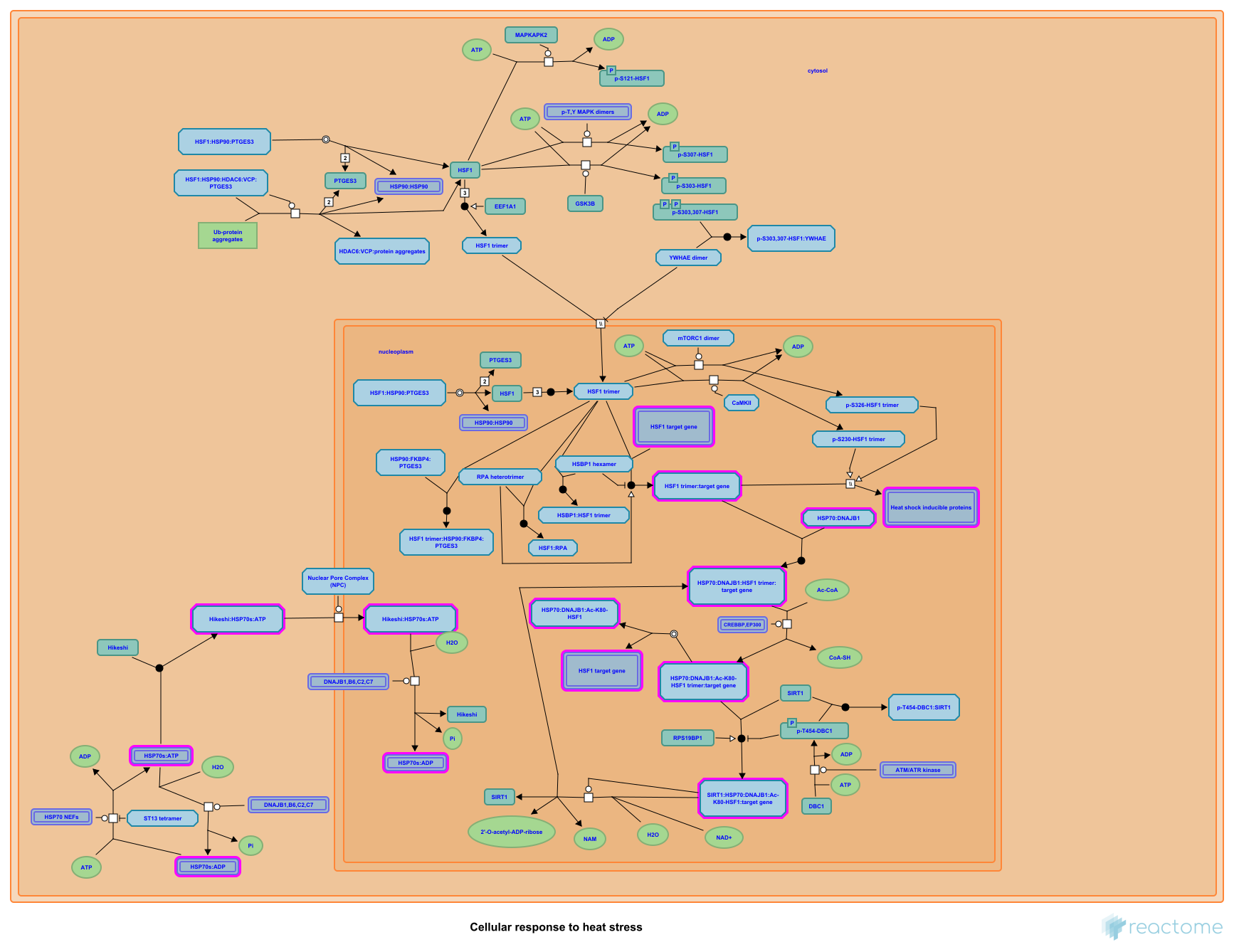
Everything in the preceding six plates happens, in a healthy cell, in seconds. HSF1 senses, HSP90 lets go, HSP70 deploys, the genome turns on the genes that make more of all three and a few dozen friends besides. The diagram above is Reactome's curated view of that cascade — R-HSA-3371556, “Cellular response to heat stress.”
The diagram is dense by design. Read it loosely — find HSPA1A (highlighted) near the centre, then trace the arrows backwards to the trimer-binding step on the DNA, and forwards to the client refolding reactions. The point of seeing it whole is that no single protein in this chapter has been operating alone. The whole network has to hold together, fast, every time the temperature climbs.
Plate VIII / 41.0 → 43.0 °C
Your environment, your enzymes
Heat as a number on a screen, and heat as a number inside a cell, are different things. The first is an environmental measurement; the second is a thermodynamic state of the molecular cast you've been reading about. The bridge between them is the body itself, mediated by hydration, acclimatization, and how long the exposure lasts.
Educational model — not a clinical prediction. The curve below is an illustrative toy: a smooth function of your three inputs, not a validated physiological simulation. No clinical decision should rest on it.
Dashed lines mark earlier-plate temperatures: filled dots appear where the projected curve crosses them.
You've reached the end of the lab. The page is a static, citation-grounded reading of a topic that, in real biology, is anything but static. If something here was wrong, file an issue; if you're working in the same space, the parent project at HeatThreshold is where the environmental safety / scheduling side lives.